The SF1 of the LBI of Osteology has two main tasks: on one hand supporting the program lines of the institute and on the other hand conducting basic and translational research.
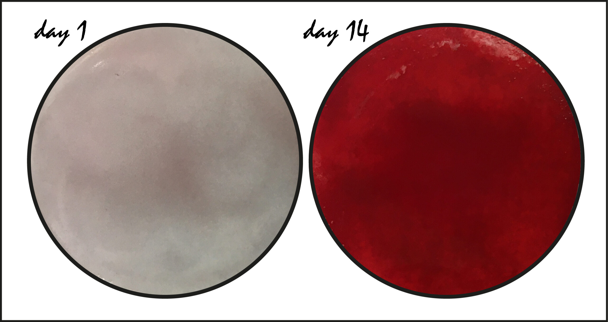
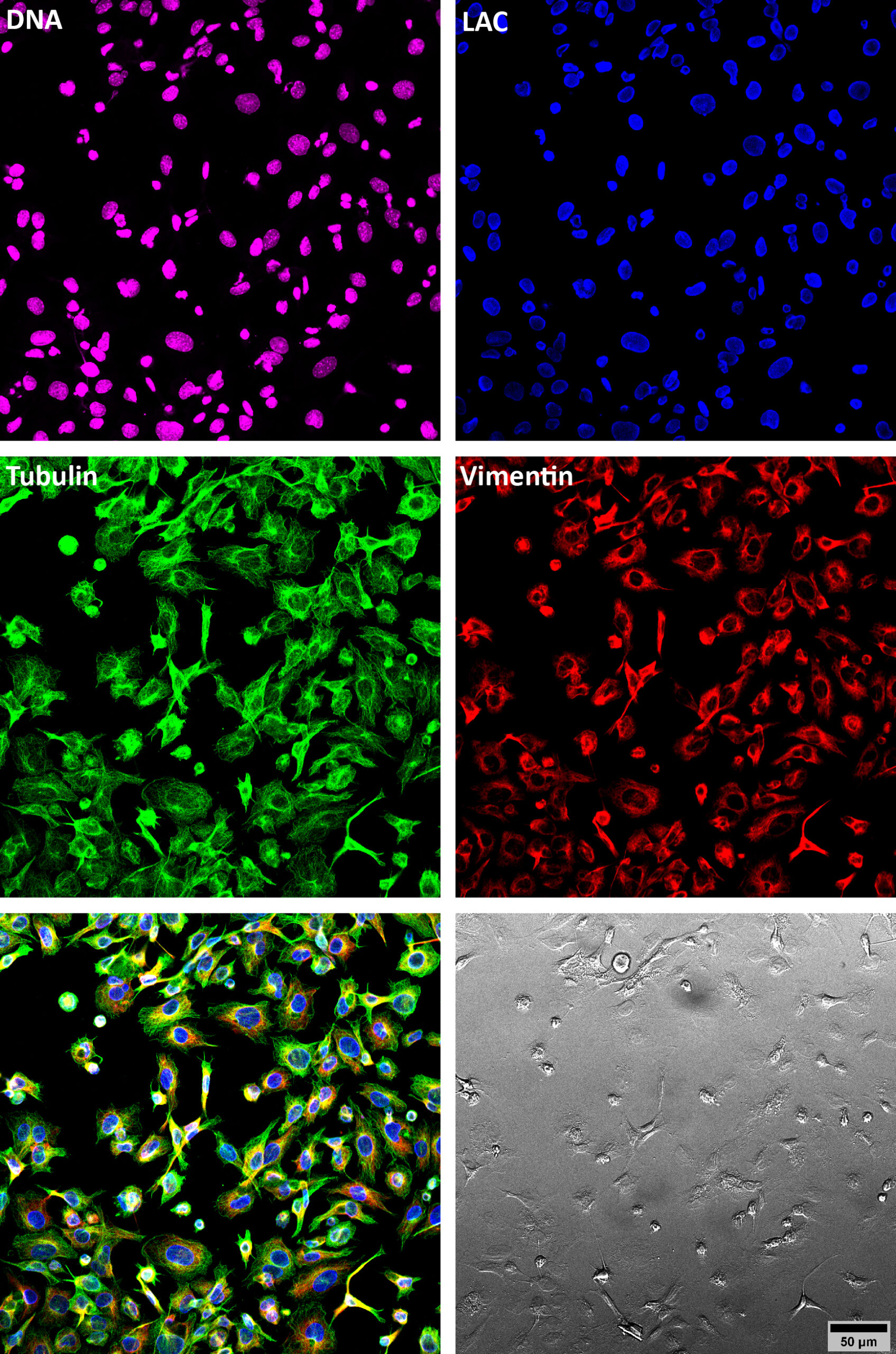
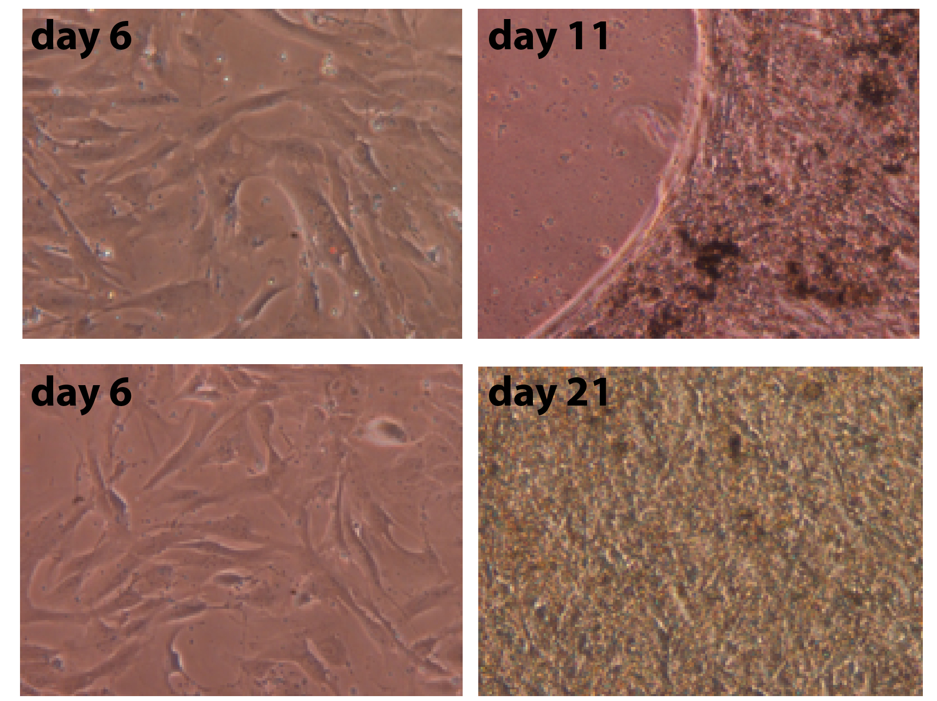
As a support facility we cultivate, expand and cryopreserve patient’s fibroblasts and osteoblasts, isolate genomic DNA for sequence analysis, coordinate bone biopsies between clinicians and the material scientists and help establishing a biobank. With respect to research we use model cell lines for bone research and employ biochemical and cell and molecular biological methods to mainly study the molecular mechanisms underlying the generation and differentiation of osteoblasts. Specifically, we are interested in the roles of the cytoskeleton, especially of intermediate filament (IF) proteins, in these processes. At the moment we focus on the type V IF proteins nuclear lamins and on the microtubule-associated motor protein kinesin-1. In another project we study the direct conversion of human dermal fibroblasts into osteoblast-like cells, a process called osteogenic transdifferentiation. In addition, in selected cases we aim to understand the impact of potential pathologic mutations identified by the Department of Medical Genetics at the Hanusch Hospital in patients exhibiting a diseased bone phenotype on cellular and molecular mechanisms using patient’s samples such as dermal fibroblasts and bone and skin biopsies.