Device
X-ray microtomography (microCT) provides non-destructive detailed 3D imaging of a wide variety of sample types. We apply microCT to numerous research problems in skeletal development and pathologies, including functional morphology and morphometric studies.
The support facility 3 has access to a new, state-of-the-art high resolution CT scanner (Scanco µCT50) allowing the scanning of ex vivo samples with a resolution up to the submicron level. It is equipped with a carrousel that loads up to 12 samples at a time.
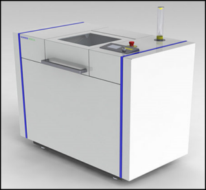
Scanco µCT50
Examples

Femur of a 20 weeks old wild-type mouse acquired with 2 µm spatial resolution

3D reconstruction of a 5×5 cm piece of bone from the human iliac bone acquired with 14 µm spatial resolution
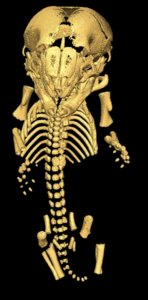
Skeletal front view of a wild type P0 mouse pup scanned with 7.4 µm spatial resolution.
Publications
Windpassinger C, Piard J, Bonnard C, Alfadhel M, Lim S, Bisteau X, Blouin S, Ali NB, Ng AYJ, Lu H, Tohari S, Talib SZA, van Hul N, Caldez MJ, Van Maldergem L, Yigit G, Kayserili H, Youssef SA, Coppola V, de Bruin A, Tessarollo L, Choi H, Rupp V, Roetzer K, Roschger P, Klaushofer K, Altmüller J, Roy S, Venkatesh B, Ganger R, Grill F, Ben Chehida F, Wollnik B, Altunoglu U, Al Kaissi A, Reversade B, Kaldis P.
CDK10 Mutations in Humans and Mice Cause Severe Growth Retardation, Spine Malformations, and Developmental Delays. Am J Hum Genet. 2017;101(3):391-403.
Fratzl-Zelman N, Roschger P, Kang H, Jha S, Roschger A, Blouin S, Deng Z, Cabral WA, Ivovic A, Katz J, Siegel RM, Klaushofer K, Fratzl P, Bhattacharyya T, Marini JC.
Melorheostotic Bone Lesions Caused by Somatic Mutations in MAP2K1 Have Deteriorated Microarchitecture and Periosteal Reaction. J Bone Miner Res. 2019 May;34(5):883-895. doi: 10.1002/jbmr.3656. Epub 2019 Jan 22. PMID: 30667555; PMCID: PMC8302214.