Material Science – Mineral, Structure, Function
Program Line 2
In PL2 we investigate various aspects of undecalcified bone matrix properties. These include the assessment of the local mineralization of bone with a spatial resolution of about one micrometer, the investigation of structural properties and cellular activity in histologically stained thin sections of bone, the representation of the osteocyte lacuno canalicular network in bone using confocal microscopy as well as the measurement of local mechanical properties using scanning acoustic microscopy. We are particularly interested in the alteration in bone material properties due to (rare) diseases.
quantitative Backscattered Electron Imaging (qBEI)
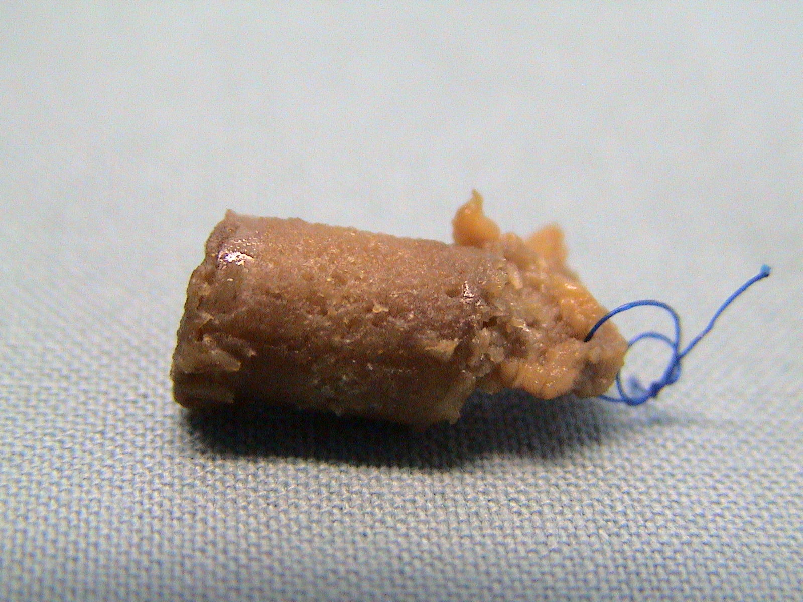
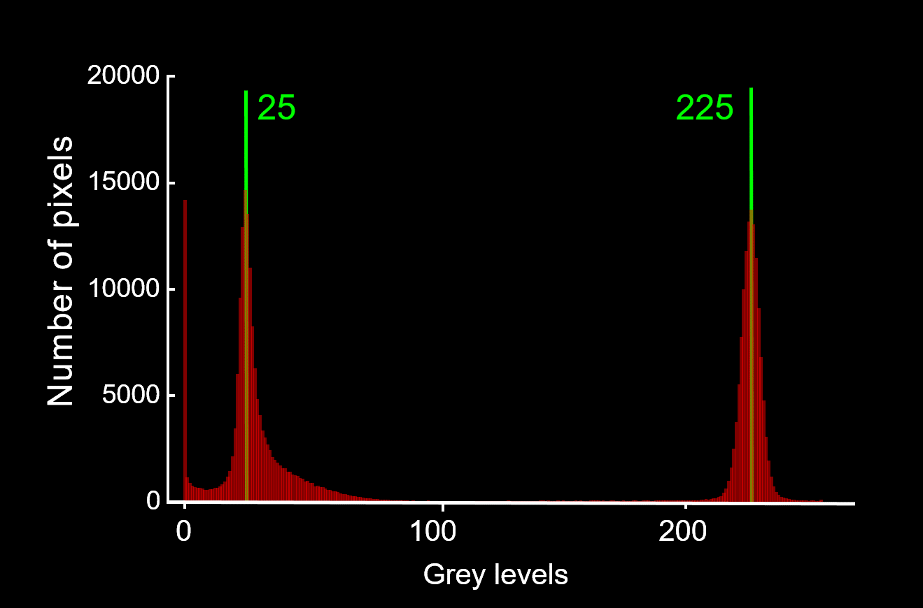
Using a scanning electron microscope it is possible to measure the local calcium content in a bone biopsy sample. In contrast to a radiographic measurement like dual energy x-ray absorptiometry (DXA), it is possible to disentangle the effect of the amount of material and mineralization of the material.
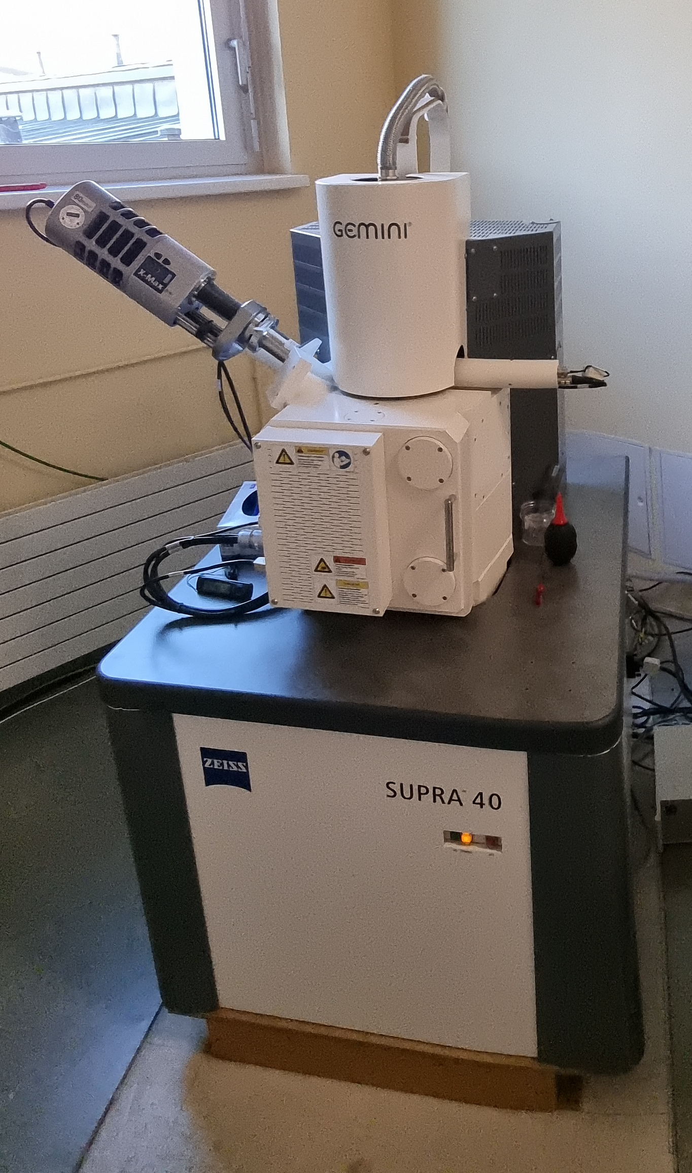
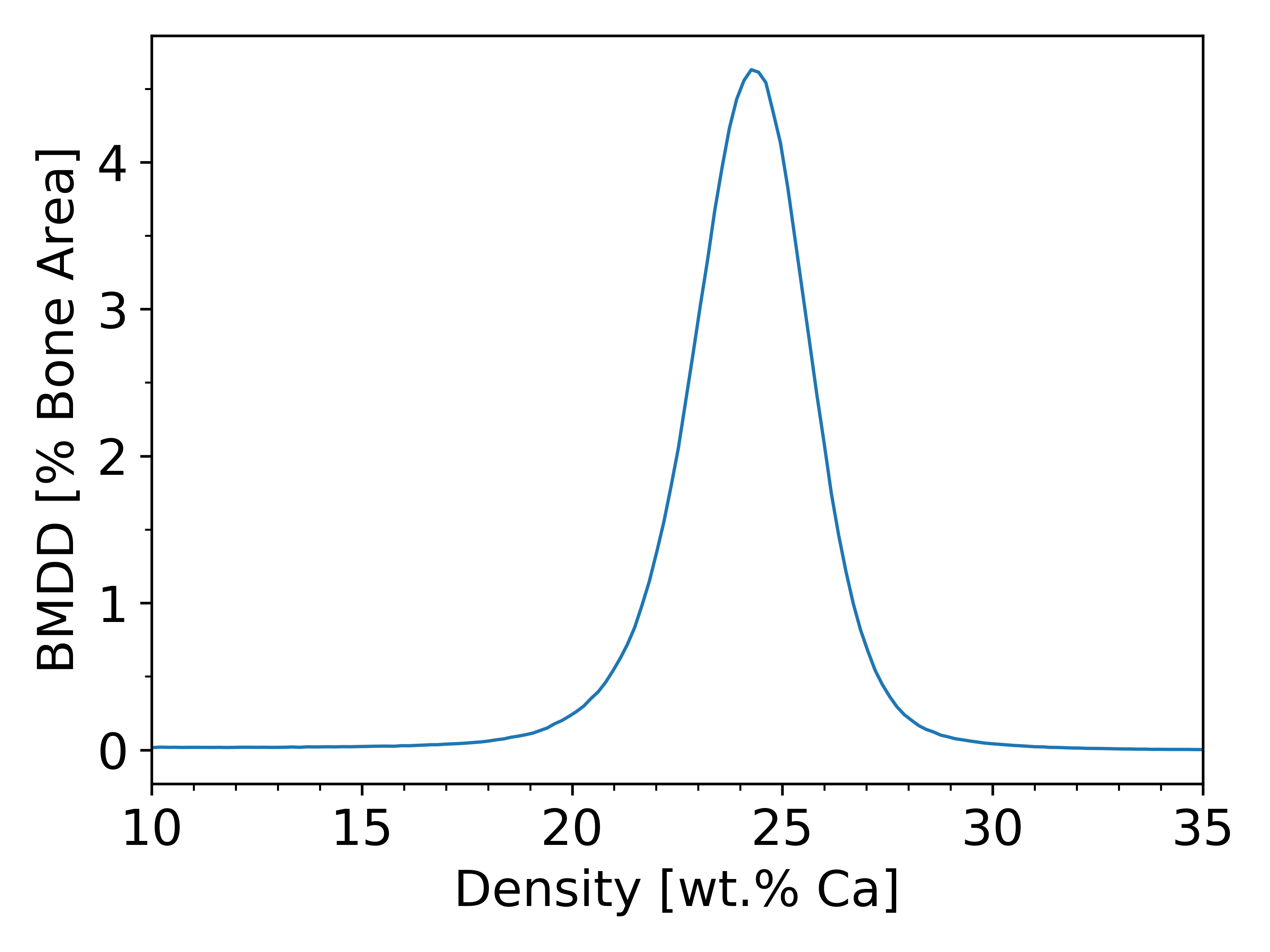
Bone is a composite material consisting of a soft, but tough organic matrix made mostly of collagen type I (85 to 90 % of total protein) as well as the hard, but brittle mineral hydroxylapatite, a calcium phosphate. Bone is constantly renewed by specialized cells, the bone forming osteoblasts and the bone resorbing osteoclasts. Osteoblasts lay down new bone in the form of unmineralized osteoid that subsequently mineralizes. This mineralization process is biphasic: during the phase of primary mineralization bone achieves around 70 % of its final mineral content within several days, while the last 30 % mineral accumulate in the phase of secondary mineralization that takes up to several months or even years. Due to the constant interplay of bone formation and resorption, bone consists of a mosaic of bone packets of different age and, thus, of different mineralization. In our group we have the possibility to measure the local mineral content (in wt.% Ca) with a spatial resolution of about one micrometer. In a scanning electron microscope (SEM) a small electron beam is scanned over the sample and the number of backscattered electrons is measured in each point. If the device is calibrated prior measurement the number of backscattered electron can be related to the local mean atomic number and, thus, to the local mineralization. Consequently, the grey level of the obtained images can be translated into a local calcium content. Subsequently, it is possible to count which amount of the bone surface is mineralized to which amount. This procedure results in the so called bone mineralization density distribution (BMDD). The importance of the BMDD stems from the observation that for adults the trabecular BMDD is independent of age, skeletal site, sex and ethnicity. Consequently, changes in the BMDD point towards a disturbed bone turnover or calcium or phospate metabolism, as is found in bone diseases like osteoporosis, osteogenesis imperfecta, Vitamin D deficiency and many others.
- Markus A. Hartmann, Stéphane Blouin, Barbara M. Misof, Nadja Fratzl-Zelman, Paul Roschger, Andrea Berzlanovich, Gerlinde M. Gruber, Peter C. Brugger, Jochen Zwerina and Peter Fratzl
Quantitative Backscattered Electron Imaging of Bone Using a Thermionic or a Field Emission Electron Source
Calcified Tissue International 109, 190 (2021) - Paul Roschger, Eleftherios P. Paschalis, Peter Fratzl and Klaus Klaushofer
Bone mineralization density distribution in health and disease
Bone 42, 456 (2008) - Paul Roschger, Himadri S. Gupta, Andrea Berzanovich, Georg Ittner, David W. Dempster, Peter Fratzl, Felicia Cosman, May Parisien, Robert Lindsay, Jeri W. Nieves and Klaus Klaushofer
Constant mineralization density distribution in cancellous human bone
Bone 32, 316 (2003)

Histological Staining and Histomorphometry
Histological staining allows to identify, distinguish and quantify different tissues (e.g., unmineralized osteoid and mineralized bone) and cells (e.g., osteoblasts and osteoclasts) to gain insight into bone metabolism and pathologies.
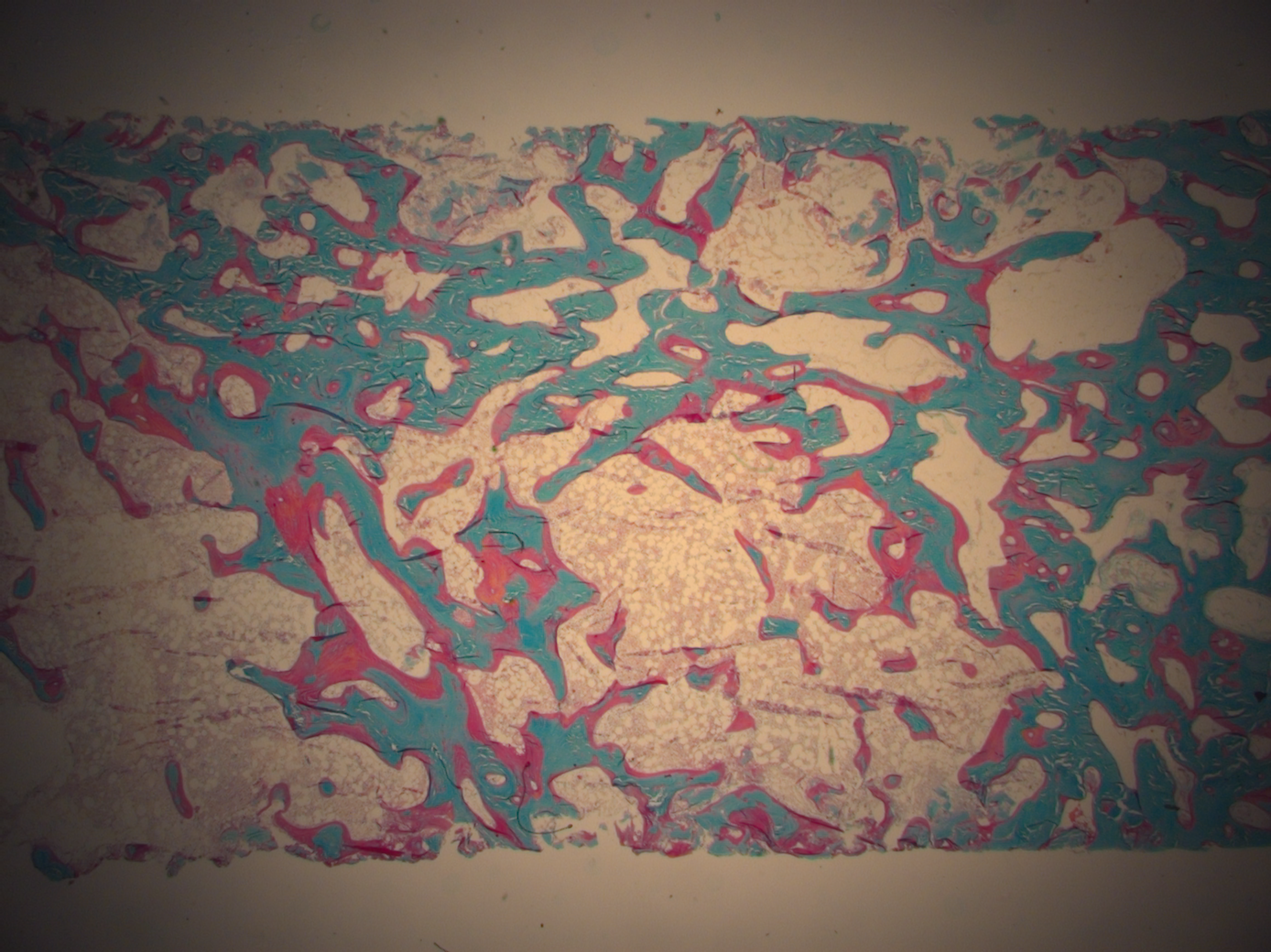
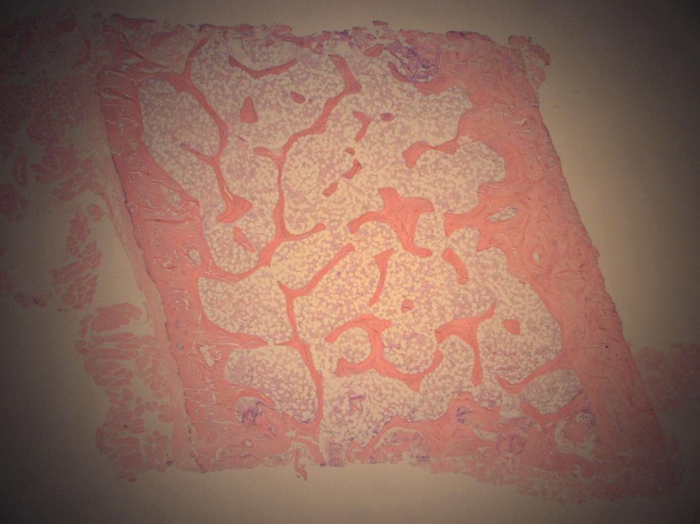
In contrast to many other labs we work on undecalcified bone samples. This means that our samples have to be plastic embedded and that special hard tissue microtomes are necessary to cut thin approximately 3 micrometer thick sections that can be further processed. In particular, we perform Goldner and Giemsa stainings on the obtained sections. Goldner stains mineralized bone in green and unmineralized osteoid in red. Thus, this staining allows to measure the relative amount of osteoid volume per bone volume, as well as other basic histomorphometric quantities like bone volume per tissue volume, trabecular number and trabecular thickness. Giemsa staining discriminates bone stained in light pink from cartilage stained in dark violett, and allows to identify bone cells like osteoblasts and osteoclasts.
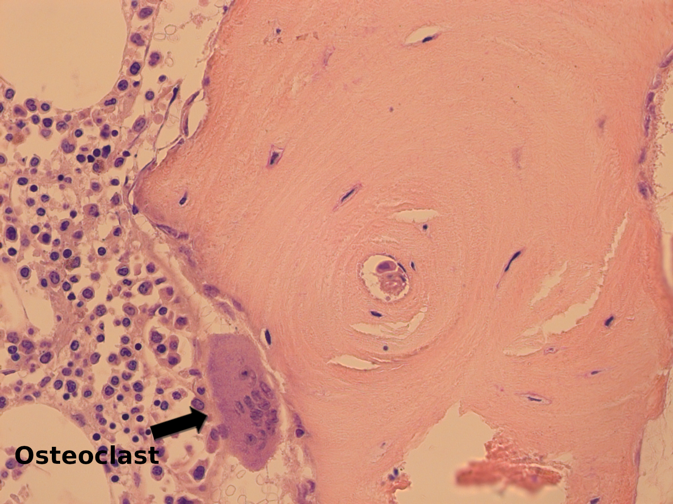
b. Giemsa staining showing a large, multinucleated osteoclast.
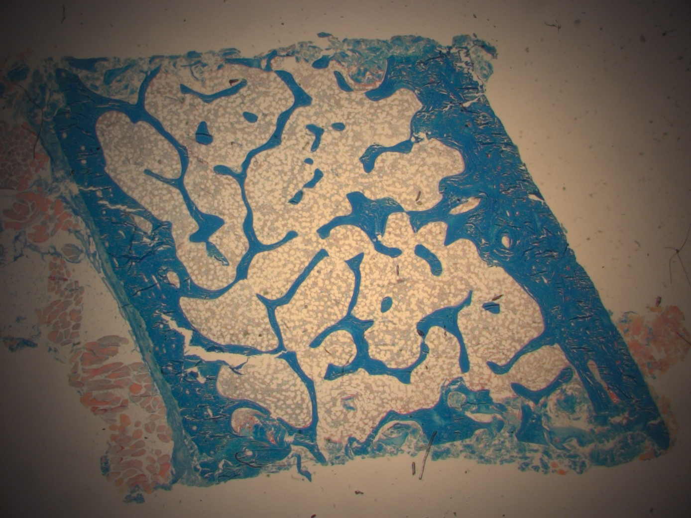
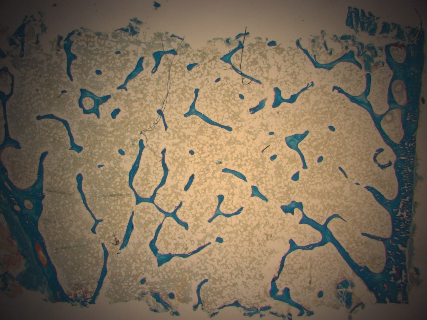
c. Goldner staining of a bone sample. Mineralized bone is stained in green, while unmineralized osteoid is stained in red.
d. Goldner stained sample from a patient suffering from osteoporosis. The reduced amount of bone material is clearly visible.
Confocal Measurements
Confocal microscopy measures fluorescent signals emerging from the sample. This gives the possibility to measure dynamic properties of bone formation (like mineral apposition rate) in labelled bone samples. As confocal measurements also allow to detect signals from emerging from different depths in the sample it also gives the possibility to map the three dimensional structure of rhodamine stained osteocyte lacuno canalicular network.
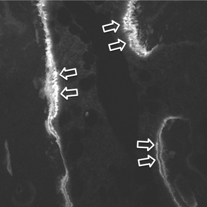
The principle of confocal microscopy is to use laser light to excite fluorescent molecules in the sample and to subsequently measure the emerging signals coming from different depths of the sample. In our group we use the confocal microsope for two different investigations. First, if possible bone samples are labelled prior biopsy with fluorescent dyes (most often tetracycline). These dyes are given twice with an interval of approximately 12 days. The dye attaches to the mineral directly at the mineralization front, thus, labelling the surface of mineral apposition at the time point of administration. Subsequently, when the sample is cut and investigated under a confocal microscope, fluorescent lines indicate the regions of mineralization. Furthermore, it can be concluded that for each double label the age of tissue in between the labels is 12 days. This procedure gives the opportunity to measure dynamic parameters of bone formation, like mineral apposition rate, mineralizing surface or bone formation rates.
Second, the confocal microscope also gives the possibility to measure the osteocyte lacuno canalicular network in 3D. Rhodamine is a fluorescent dye that stains all accessible bone surfaces, in particular also the osteocyte lacunae and the canaliculi. Consequently, using the confocal microsope it is possible to record the signal coming from rhodamine for various depths in the sample. The obtained image stack can then be used to reconstruct the network in full 3D.
- Felix Repp, Philip Kollmannsberger, Andreas Roschger, Michael Kerschnitzki, Andrea Berzlanovich, Gerlinde M. Gruber, Paul Roschger, Wolfgang Wagermaier and Richard Weinkamer
Spatial heterogeneity in the canalicular density of the osteocyte network in human osteons
Bone Reports 6, 101 (2017)
The osteocyte lacuno canalicular network in a mouse visualized with confocal microscopy.
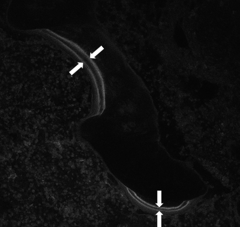


b. A large amount of smeared double lines indicates a mineralization defect, possibly an osteomalacia.
c. The osteocyte lacuno canalicular network in bone measured with confocal microscopy using rhodamine stained samples.
d. Full 3D structure of the osteocyte lacuno canalicular network
Rare Diseases
Rare Diseases affect less than 1 in 10000 people. We are interested in the alterations of bone material quality and quantity that may go along with such pathologies and that are often the cause of frequent fractures or deformities.
Rare diseases investigated in our group are Osteogenesis Imperfecta, pycnodysostis, x-linked hypophosphatemia (XLH) or melorheostosis. As bone biopsy samples from such patients are naturally rare, it is important to investigate the available samples with all possible methods to get most information out. In some cases it is also possible to investigate a mouse model carrying the same mutation as the human counterpart.
- Matthias Mähr, Stéphane Blouin, Martina Behanova, Barbara M. Misof, Francis H. Glorieux, Jochen Zwerina, Frank Rauch, Markus A. Hartmann and Nadja Fratzl-Zelman
Increased Osteocyte Lacunae Density in the Hypermineralized Bone Matrix of Children with Osteogenesis Imperfecta Type I
International Journal of Molecular Sciences 22, 4508 (2021) - Nadja Fratzl-Zelman, Sonja Gamsjaeger, Stéphane Blouin, Roland Kocijan, Pia Plasenzotti, Stamatia Rokidi, Kamilla Nawrot-Wawrzyniak, Katharina Roetzer, Gökhan Uyanik, Gabriele Haeusler, Elizabeth Shane, Adi Cohen, Klaus Klaushofer, Eleftherios P. Paschalis, Paul Roschger, Peter Fratzl, Jochen Zwerina and Elisabeth Zwettler
Alterations of bone material properties in adult patients with X-linked hypophosphatemia (XLH)
Journal of Structural Biology 211, 107556 (2020) - Nadja Fratzl-Zelman, Paul Roschger, Heeseog Kang, Smita Jha, Andreas Roschger, Stéphane Blouin, Zuoming Deng, Wayne A. Cabral, Aleksandra Ivovic, James Katz, Richard M. Siegel, Klaus Klaushofer, Peter Fratzl, Timothy Bhattacharyya and Joan C. Marini
Melorheostotic Bone Lesions Caused by Somatic Mutations in MAP2K1 Have Deteriorated Microarchitecture and Periosteal Reaction
Journal of Bone and Mineral Research 34 883 (2019) - Nadja Fratzl-Zelman, Angelika Valenta, Paul Roschger, Alexander Nader, Bruce D. Gelb, Peter Fratzl and Klaus Klaushofer
Decreased Bone Turnover and Deterioration of Bone Structure in Two Cases of Pycnodysostosis
Journal of Clinical Endocrinology and Metabolism 89, 1538 (2004)